Nora is a web-based framework for medical image analysis. It has been developed to bridge the gap between research and clinic, and to boost medical imaging research to the next level. It provides a high-level web-interface accessible from any webbrowser to visualize, organise, process and share data in a very customizable way.
Depending on your needs, your Nora instance can run as a web-service in the cloud or as a local installation at your institution.

Generic placeholder image

Visualization

Use the nora viewer to explore and interact with your data. Use smart loaders to view data with pre-defined configurations.

Generic placeholder image

Processing Platform

Collect, manage, and share multimodal data from different folders, harddrives or PACS databases.
Process the data using your own functions, or existing toolboxes.

Generic placeholder image

Reading / Labeling

Use the power of the web-based tools to perform interactive reading studies, and label, annotate and segment ground truth datasets.

Generic placeholder image

CAD reports

Provide and get access to cutting-edge computer aided diagnosis methods with automated workflow from scanner to report.


nora viewer
nora viewer Runs directly in any webbrowser.

Nora medical image viewer. Not only for researchers.

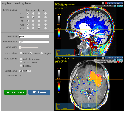
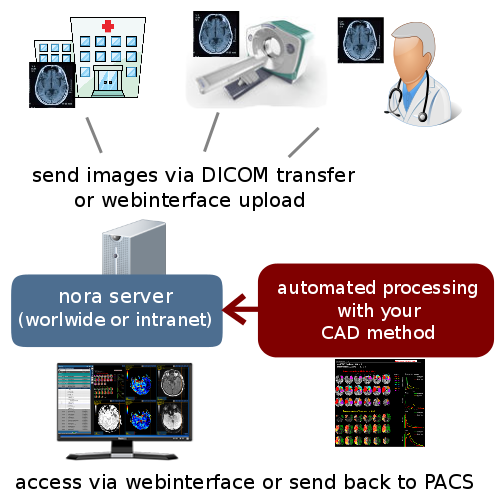
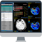
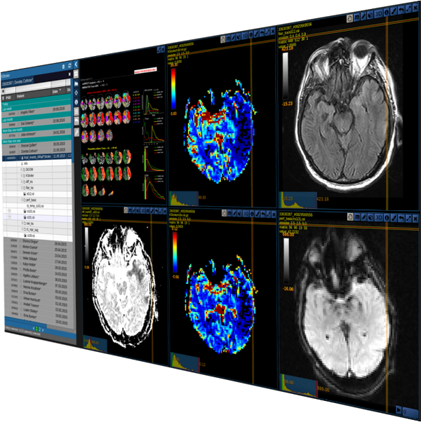
The Nora image viewer runs in any web-browser, without installation or update issues. It provides features beyond the standard, such as real-time image resclicing (MPR), overlays, ROIs, 3D surface rendering and connectome/fiber viewer.
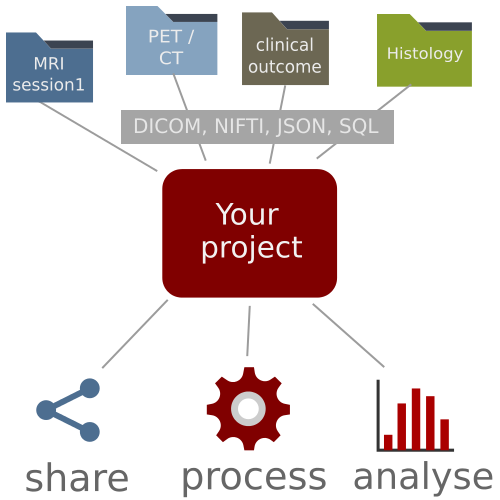
Besides medical image file formats like DICOM, NIFTI and BRUKER, the viewer has functionalities to show other common formats like json, jpeg, png, pdf. With this, you will always have all relevant data at one glance. 
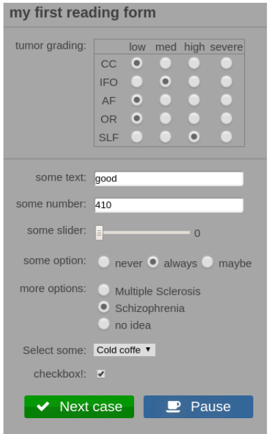
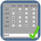
The viewer is highly customizable: For different projects, it can be configured with preset schemes to automatically load specific datasets with specific settings.
The viewer also works as a standalone offline tool: Without installation, simply drag and drop your local files into the browser and use all of the useful features.

Learn more

Back to top

Generic placeholder image
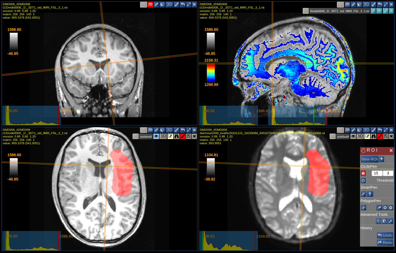
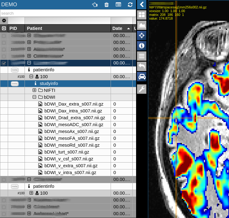
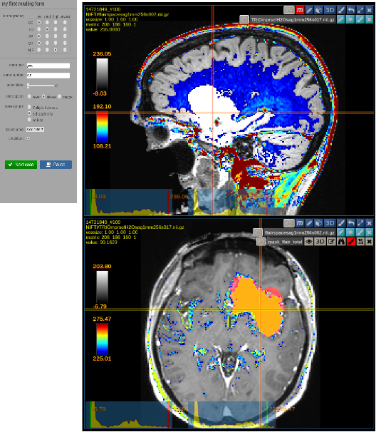
Image viewer: All standard features are available, such as correct interpretation of 3D orientation information, real-time reslicing (MPR reconstruction), slicing of different images in same voxel space, overlays and rois.
Generic placeholder image
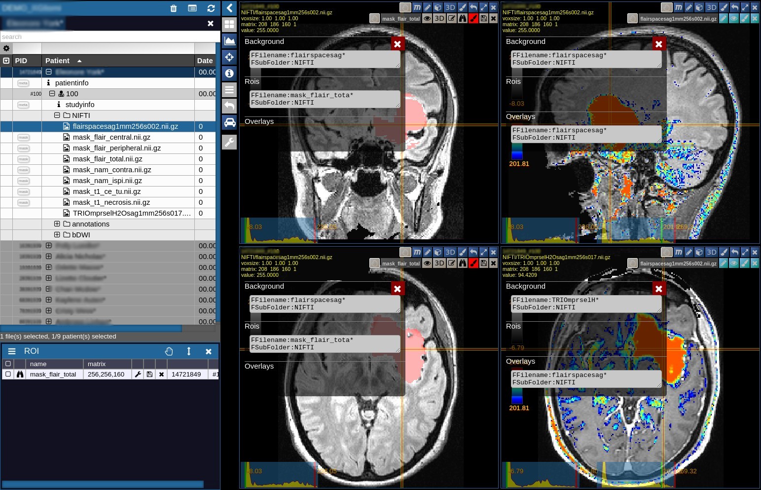
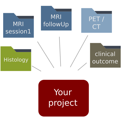
Smartloader: Define which images / metadata you want to automatically load when selecting a patient from your project, for example "Load the flair MRI scan as an orthoview and overlay with a PET image from another session", or "In a longitudinal study, load all available flair images for each visit in different sub-windows."
Generic placeholder image

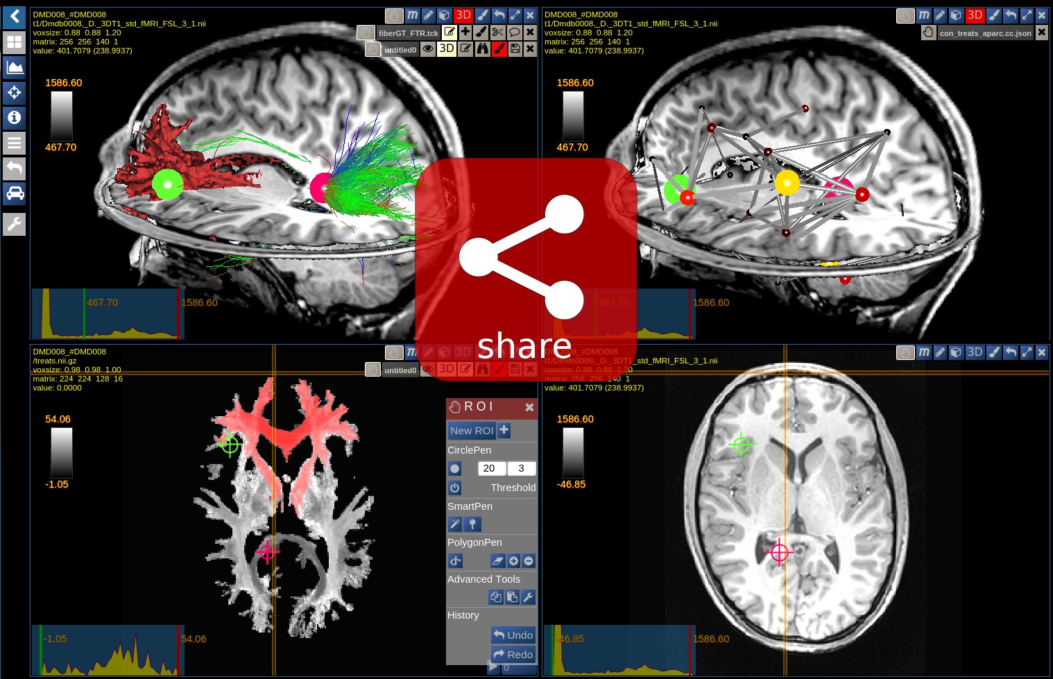
Smartlink: Create an "interactive snapshot" of your current view and send a shared link to your colleague. In his webbrowser, the exact same configuratin will be displayed.
Generic placeholder image

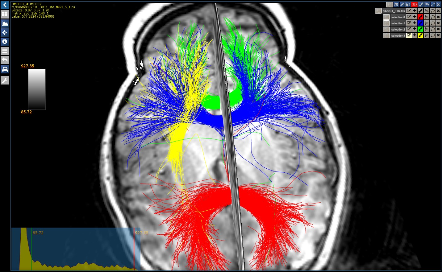
MRI Tractogram Fibers: The fiber viewer provides features beyond the standard: Navigate in 3D and follow tracts interactivley and in real time using the optimized octtree functionality.
Generic placeholder image

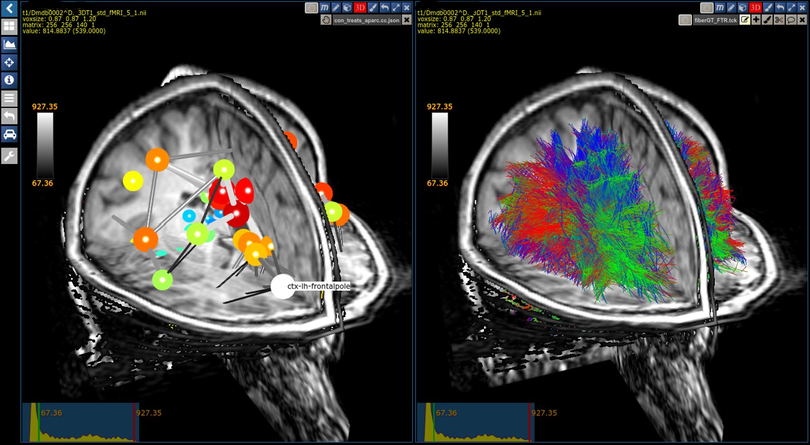
MRI Tractogram Connectivities: Navigate through the brain and explore the connectivites.
Generic placeholder image

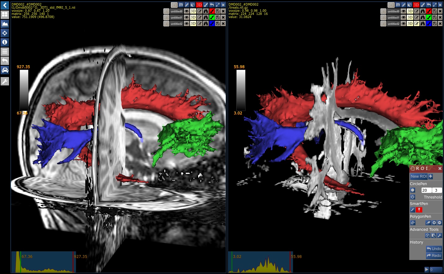
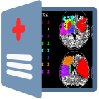
MRI Tractogram Segmentations: Interactivley segment tracts in real time based on a machine learning algorithm.

Generic placeholder image

The Nora processing platform.
Manage, process, analyse.

Manage, visualise, process and analyse your images on a web-based multi-user platform. Include data from various inhomogeneous sources, no matter if file-based, spread over multiple harddrives, or imported from a PACS system. In the webinterface, you will always have all data at one glance.
For image processing, Nora provides a unified interface. Use existing toolboxes such as SPM, FSL or FreeSurfer and others, without dealing with their individual function calls, or implement your own algorithms with python, matlab or as command line tool. You can simply define your batch pipelines in the webinterface, and start processing hundreds of patients on a parallel grid engine with a single mouseclick. Finally, retrieve a results table with calculated quantitative parameters in selected regions of interest chosen from built-in atlases or segmented manually.

Learn more

Back to top

Generic placeholder image

1. Nora Data Management.
Be organised.

With Nora, you can effectively join all your data for a specific project. No matter how your data is distributed, simply add your data folders to your project, Nora will gather all information in a database. The Nora data management can deal with any data folder organisation, as long as some ID can be extracted from the directory names. This way, you can join data from many different locations and store calculated results in another folder structure or location, without loosing track.

You can also add remote data from mounted network drives or cloud storage. The Nora database only holds meta information, the data itself always remains where it is. The system also provides a DICOM interface to exchange data with other PACS systems or imaging devices.

Generic placeholder image

2. Nora Patient Browser.
Everything at one glance.

No matter how your data is distributed, in the web-interface everything will show up well organised at one glance, searchable, browsable and sharable to colleagues working on this project. Here, you can also set up your SMARTLOADERS to define which images should be loaded in which configuration when selecting a patient.

A new Patient, sent from a PACS, uploaded via the webinterface or mounted as a new network drive will automatically pop up in the list. You can set tags, add metadata, browse, sort, search, define groups and and start the data processing from here.

Generic placeholder image

3. Nora data processing.
One interface, all toolboxes.

Nora provides a high level interface for existing imaging processing toolboxes, or your own code. No matter if you want to process data with your matlab code, command line tools or toolboxes like SPM, FSL or FreeSurfer: Don't care about the specific function calls of the different programming languages and toolboxes.

Define your calculation pipeline via the webinterface and process thousands of datasets with a single mouseclick.

A grid engine will take care for parallel computing of your jobs, with full control over optimal workload, job status, progress and errors.

Generic placeholder image

4. Nora statistics.
Aggregate your result.

In many cases, data from clinical trials is evaluated based on statistical measures. These can be general global parameters, such as clinical scores, or image-derived parameters from functional MRI, diffusion or perfusion imaging, or any other quantitative measure. In many cases, these values are to be compared in selected regions of interest.

With a single mouseclick, you can aggregate these values for huge clinical trials and retrieve a table of results, joined with any other metadata.


Generic placeholder image

Nora reading / labeling.
Create your groundtruth.

Nora allows for interactive reading, labeling and tagging in a multi-user environment. You want to investigate whether prostate cancer can better be assessed using a combination of PET/CT or DCE MRI? Or you want to know if your automated segmentation algorithm really can support visual diagnosis of lung cancer?
Set up a project and define an interactive form with click, check, and combo-boxes. Invite your colleagues worldwide to read and rate the cases in a randomized manner and / or in multiple stages showing the data in different configurations. With this standardisation, you can get results with great statistical power.
This is not only a powerful tool for randomized surveys, but also to perform quality checks and to generate ground truth datasets with labels, annotations and regions of interest.

Learn more

Back to top

Generic placeholder image

1. Create your questionnaire.

Define your questionnaire using elements such as
  • checkboxes
  • choice grids
  • sliders
  • free text ...
You can also introduce dependencies such as "if option A was chosen, show another sub-box with a refining question. "
Generic placeholder image

2. Configure your view.

With the SMARTLOADER of the viewer, you configure what to show for each case
  • which images in which viewport
  • colormaps and default windowing
  • overlays, rois
  • additional patient information and metadata such as clinical scores


Configure the reading in terms of
  • randomisation order
  • repetition of cases
  • multiple stages
  • what to show in each stage
  • let readers interactively draw segmentations or annotations
  • record additional parameters such as time spent per case, click behaviour
Generic placeholder image

3. Run your reading study.

Invite readers from your intranet or from all over the world to participate in your reading study. No matter if they sit in their office in New York, or at the airport in Shanghai, they can walk through the cases from anywhere in the world, whenever they want.

The system is easily scalable: Once your reading is set up, it doesn't matter how many readers access the system. With this, your statistical significance will increase enormously, and you can create very reliable ground truth datasets.

You can always follow the current status of the reading. Once all readers have done their job, download the results table and publish your high impact paper.


Generic placeholder image

Nora CAD.
Instant Computer Aided Diagnosis.

Applications with fully automated processing pipelines can provide very valuable support for clinical diagnosis. Especially the emerging field of machine learning methods, based on the combination of multimodal data is regarded as an important tool for improved personalised healthcare.
With Nora, scientists, software engineers and manufacturers can provide their applications to a large community in order to test and improve their CAD methods. Clinicians all over the world, not only from large clinical centers but also from smaller hospitals can gain access to cutting edge research technologies.

Learn more

Back to top

Generic placeholder image

1. Computer Aided Diagnosis.
Everything at one Glance.

The usecases of computer aided diagnosis are manifold. No matter if automated detection of lesions, tumor segmentations or the combination of multimodal data (e.g. MRI and PET, genomics, other clinical parameters), they can add very valuable diagnostic value, especially in the context of machine learning.

Diagnostic reports also have the power to improve standarisation in medical imaging: They provide clinicians always with the same clear and standardised view of all relevant images and derived parameters.
Generic placeholder image

2. Bridge the gap.
It's all about infrastructure

Each year, researchers are producing and improving dozens of potentially valuable medical image processing methods. However, the transfer from research to clinical practice leaves much to be desired.

One reason is the lack of an infrastructure to easily and quickly provide, evaluate and optimise prototypes in a clinical environment.

Even though many applications might work "in principle", a seamless workflow regarding image acquisiton, data transfer, data processing and visualisation is a crucial to provide methods to clinicians.
Generic placeholder image

3. Bring research to the clinic.

Nora can provide such a platform-independent infrastructural framework.

Implement your code in any programming language, access them with noras high-level processing interface and let computational intense calculations automatically run in parallel on a server farm. This way, you can easily provide and test your research methods in a clinical setting.
First, restricted to your local institution, and later to clinicians all over the world.

They can send their images via the DICOM transfer protocols, or as upload in the webinterface. Images will automatically be processd and can be accessed via the webinterface, or sent back to the users PACS.

Finally, the feedback of applicants helps you to evaluate and optimise your algorithms.

Downloads Generic placeholder image

To get an impression of NORA's viewing capabilities you can download here parts of NORA's viewer packaged within the Electron framework as an ordinary desktop application. The viewer can read various medical/scientific imaging formats including DICOM, nifti, mgh, tck, trk, Bruker, gifti, stl


Who we are

Nora is developed by a team of researchers and clinicians at the University Medical Center Freiburg, Germany.

Names below are in given in alphabetical order

Project Lead:

Dr. Elias Kellner

Dr. Marco Reisert

Clinical Lead:

Dr. Maximilian Russe

Project Mentors:

Prof. Dr. Dr. Fabian Bamberg

Prof. Dr. Volker Coenen

Prof. Dr. Dr. h.c. Jürgen Hennig

Prof. Dr. Horst Urbach

Clinical Partners:

PD Dr. Jonas Hosp

Prof. Dr. Elmar Kotter

Prof. Dr. Marc Christian Metzger

Dr. Alexander Rau

PD Dr. Christopher Schlett

Prof. Dr. Cornelius Weiller

Scientific Partners:

PD Dr. Valerij G. Kiselev

Dr. Henrik Skibbe

Generic placeholder image

Contact:
NORA Medical Imaging Platform Project
Dr. Elias Kellner
Dr. Marco Reisert
University Medical Center Freiburg
Department of Radiology
Medical Physics
Killianstr. 5a
79106 Freiburg, Germany

+49 761 270 93860
elias.kellner[at]uniklinik-freiburg.de
marco.reisert[at]uniklinik-freiburg.de


Back to top

You want to see a live demo or start a project with us? Just Drop us a line!